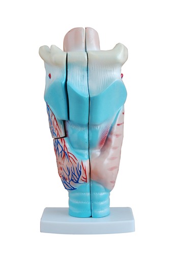
Anatomical Human Enlarged Larynx Model
This model is a visual aid, ideal for teaching the larynx structure in schools and colleges.
It helps the students to acquire an understanding of the morphology and structure of the respiratory tract and the organ of speech (phonetic organ).
It demonstrates the thyroid cartilage, cricord cartilage, epiglottis, laryngeal muscles as well as a part of the ligaments that make up the larynx. The thyroid gland is shown by its left and right lobes.
On the median sagittal section, the model shows the false vocal cord (plica ventriculus), true vocal cord (plica phoneticus) and laryngeal ventricle etc. of the phonetic organ. Above and anterior to the larynx is the hyoid bone, while the lower part of the larynx is connected with the trachea.
The model is dissectible into 3 parts and is made of washable, high-quality PVC. It is 3 times enlarged in size.
Size: 23x12.5x26.5cm
